Cherry eye in dogs, that pink bulge, what it is, how to fix it
Cherry eye is a common, distinctive eye condition in young dogs of certain breeds, a sudden pink or red bulge in the corner of the eye that wasn't there yesterday. It looks alarming. It's not life-threatening. Below: what it actually is, why surgery is the answer, what it costs in Australia, and why English Bulldogs are basically the universe's official cherry-eye sponsor.
What cherry eye actually is
Dogs have three eyelids, upper, lower, and a third eyelid in the inner corner of the eye called the nictitating membrane. The third eyelid contains a small tear-producing gland that's normally tucked out of sight. When the ligament holding this gland in place is weak or torn, the gland prolapses, pops out of position, and shows as a pink or red bulge in the inner corner of the eye.
That's cherry eye. The "cherry" name comes from the rounded, red, shiny appearance. (Whoever named it was either hungry or unkind.)
What cherry eye looks like
Cherry eye is unmistakable once you've seen it:
- A pink, red or salmon-coloured bulge sitting in the inner corner of the eye, near the nose
- Usually round to oval, varying from pea-sized to almost half the visible eye area
- Smooth, shiny, sometimes slightly glistening
- Appears suddenly, often overnight
- Affects one eye initially, sometimes both eyes within months
- Can come and go in early stages, going back into position then popping out again
- Doesn't usually cause obvious pain, though some dogs paw at the eye or rub it on the floor
Cherry eye is sometimes confused with conjunctivitis or eye infection. The key difference is that cherry eye is a discrete round bulge, not generalised redness. If unsure, see a vet.
Which breeds are prone to cherry eye?
Some breeds are far more likely to develop cherry eye due to genetic ligament weakness:
- English Bulldogs and French Bulldogs
- Beagles
- Cocker Spaniels
- Cavalier King Charles Spaniels
- Lhasa Apsos and Shih Tzus
- Boston Terriers
- Mastiffs and Bullmastiffs
- Saint Bernards
- Cane Corsos
Most cherry eye cases occur in dogs under 2 years old, often in the first 6 to 12 months of life. Mixed breeds with these breeds in their background can also develop it.
Causes
The main cause is a weak or torn fibrous attachment between the third eyelid gland and the orbital tissue. Mostly genetic, which is why specific breeds dominate cases. Trauma, allergies and irritation can sometimes trigger a prolapse in a predisposed dog.
The third eyelid gland is responsible for around 30% of total tear production. Long-standing prolapse can affect the gland and reduce tear output. That's why prompt treatment matters.
Can cherry eye go away on its own?
Usually no. Some early cases pop in and out for a while before settling permanently in the prolapsed position. Spontaneous, lasting resolution is rare. Treatment is needed in most cases.
Some vets will try gentle massage of the gland back into position with topical lubricants and anti-inflammatory drops as a first-line approach in very early cases. Sometimes works for hours or days, but recurrence is the rule.
Treatment options
Surgical tucking (the preferred approach)
The gold standard is surgical replacement of the gland, the gland is tucked back into its proper position and held there with internal sutures or a "pocket" technique. The gland is preserved, tear production continues normally, and the cosmetic result is good.
Modern Australian vet ophthalmologists overwhelmingly favour gland-saving surgical techniques over removal. Success rate is around 80 to 90% for first-time procedures, with some recurrences requiring revision surgery.
Surgical removal (last resort)
In the past, the prolapsed gland was simply removed. This is now considered a last-resort option because removing the gland predisposes the dog to lifelong dry eye (keratoconjunctivitis sicca, or KCS), which requires daily eye drops for life. Most modern vets won't remove the gland unless other approaches have failed and the gland is severely damaged.
Medical management
Topical anti-inflammatory and lubricating drops can manage symptoms while you wait for surgery, or for very mild intermittent cases. They don't cure cherry eye but can reduce inflammation and discomfort.
Cherry eye surgery cost in Australia
Specialist ophthalmologists are the safer bet for breeds with high recurrence risk and for second-eye cases. The price difference is usually justified by lower recurrence rates and better cosmetic outcomes. If cost is a barrier, the vet payment plans guide covers options.
Recovery after cherry eye surgery
Recovery is typically straightforward:
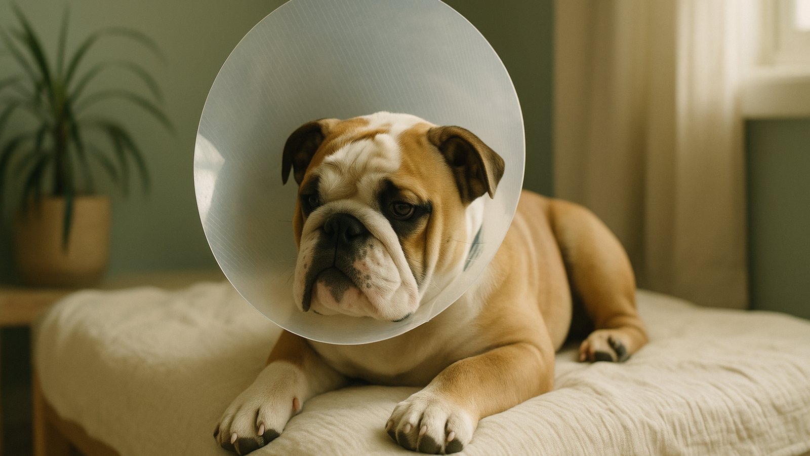
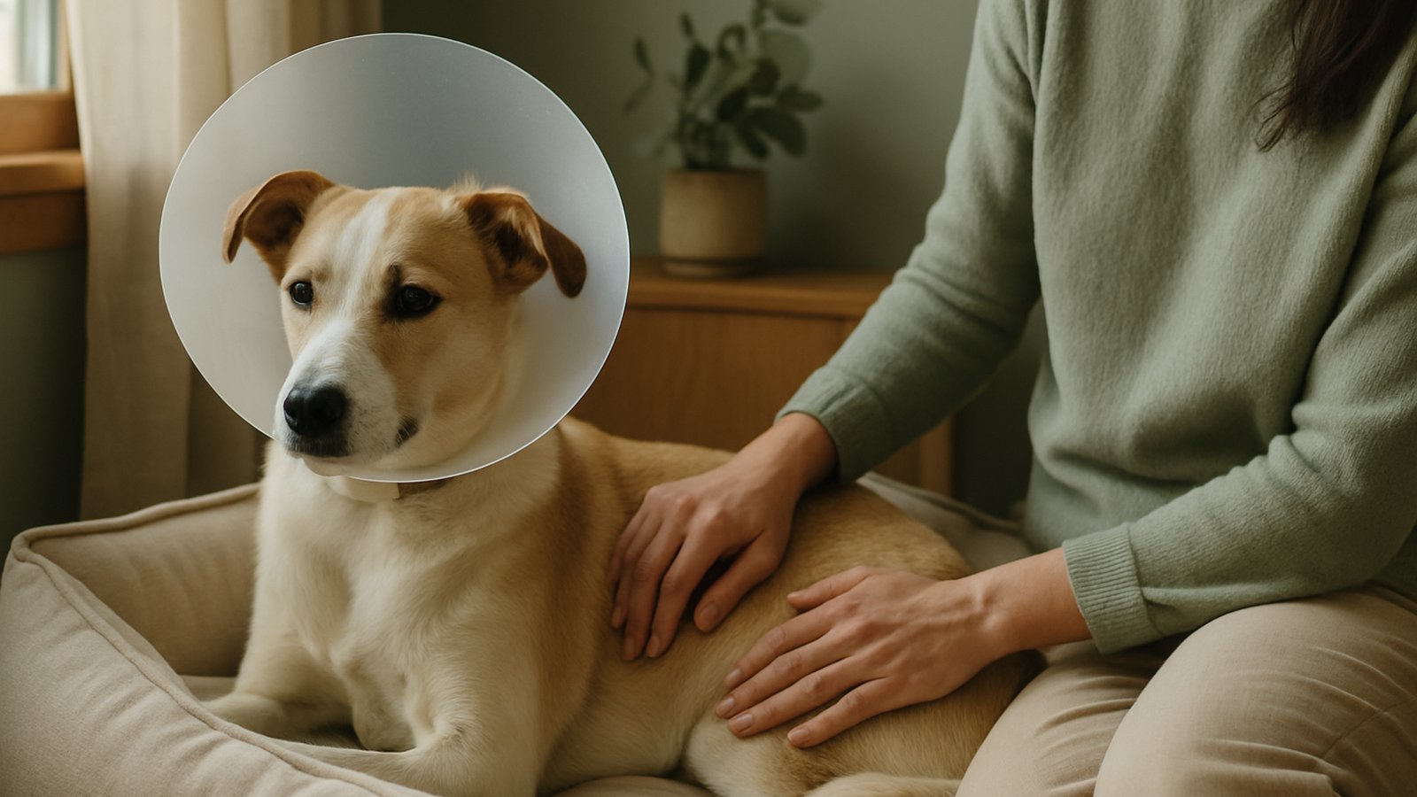
- Day 1. Sleepy from anaesthesia, mild eye swelling, sometimes red and watery. Cone on at all times to prevent rubbing.
- Days 2 to 7. Topical eye drops 2 to 4 times daily. Reduced activity. Cone stays on. Some dogs are nearly back to normal at this point; others have visible swelling for a couple of weeks.
- Days 7 to 14. Suture check or recheck appointment. Cone usually still on.
- 2 to 4 weeks. Inflammation resolves. Eye looks normal. Cone comes off after vet clearance. (Your dog has been wearing a satellite dish for two weeks. Be gracious.)
Warning signs after surgery
- Worsening swelling after day 3
- Yellow or green discharge
- The eye held closed or squinting heavily
- The cherry eye reappearing
- Aggressive head shaking or pawing despite the cone
Recurrence is most likely in the first weeks. Strict cone use and following post-op instructions matters. If you suspect recurrence, see your vet, early revision is usually simpler than late revision. For other eye-related issues that need urgent care, see the emergency vet guide. Need help finding a clinic? The find a vet guide has the run-down. (Cherry eye also tends to follow the same brachycephalic breeds prone to grooming-related skin fold issues, same dogs, different problems.)
Cherry eye is one of those things that looks much worse than it is. Surgery, two weeks in a cone, and your dog forgets it ever happened. You'll forget too, eventually. Information here is general; cherry eye should always be assessed and treated by a registered veterinarian.